The technology from Cancer Grand Challenges team IMAXT uses advanced spatial biology techniques to analyse tumours, some of which are based on technology originally developed to map the Milky Way and discover new planets. Now, other scientists will be able to access these technologies to create detailed tumour maps that could one day transform how we diagnose and treat cancer.
Led by Professor Greg Hannon and Dr Dario Bressan at the Cancer Research UK Cambridge Institute and Dr Nicholas Walton at the University of Cambridge’s Institute of Astronomy, SPACE will give other researchers the opportunity to study cancer in a way that wasn’t previously possible.
Dr Dario Bressan, Head of the SPACE Laboratory at the Cancer Research UK Cambridge Institute, said: “Tumours aren’t just a uniform mass of cells; they consist of a diverse ecosystem of cancer cells, immune cells, and other essential components that support their survival. Hidden within these intricate networks lies valuable information which could guide us in making more personalised treatment decisions for each patient.
“With the SPACE platform, researchers can zoom into specific cell populations, highlight the complex connections between them, and even run virtual experiments to predict how the tumour might respond to different treatments. By unlocking these insights, we can transform the future of cancer care and uncover new opportunities for targeted therapies.”
The IMAXT team was first awarded £20 million in 2017 by Cancer Research UK through Cancer Grand Challenges, a global research initiative co-founded by Cancer Research UK and the National Cancer Institute in the US.
Since then, the team has united experts from fields rarely brought together including medicine, Virtual Reality (VR), programming, molecular biology, chemistry, mathematics, and even astronomy, to create a completely immersive tool for studying tumours.
As well as enabling scientists to analyse 3D tumour maps, IMAXT created pioneering VR technology which allows the user to ‘step inside’ a tumour using a VR headset.
With the headset, scientists get to view vast amounts of detailed data about individual tumour cells in a 3D space. Instead of looking at this data on a computer screen, they can see all the information in real-time, as if they were inside the tumour itself.
Professor Greg Hannon, Director of the Cancer Research UK Cambridge Institute, said: “Cancer Grand Challenges offers a unique opportunity for international teams to address some of cancer’s biggest challenges. When we took on our particular challenge, much of what we proposed was science fiction.
“Over the past 7 years, our team has turned those early hopes and ideas into approaches that can now be made broadly available. In nature, biology unfolds in three dimensions, and we now finally have the tools to observe it that way—giving us a much deeper, more accurate view of cancer. We’re thrilled to share these breakthroughs with the broader cancer research community.”
Director of Cancer Grand Challenges at Cancer Research UK, Dr David Scott, said: “IMAXT is changing what’s possible when it comes to cancer research.
“We can glean important insights about a tumour by analysing its genetic makeup or its proteins, but no technology alone can give us the depth of understanding needed to truly understand this complex disease.
“By combining state-of-the-art technology and vast expertise, IMAXT will change how cancers are classified, treated and managed, giving more people a better chance of surviving their disease.”
The funding will support the SPACE hub laboratory, hosted at the CRUK Cambridge Institute, and the SPACE analysis and computing platform, developed and operated at the Institute of Astronomy, University of Cambridge. Together SPACE includes and combines most available technologies for the spatial molecular profiling of tumours. The continued collaboration between the cancer and astronomy teams from the IMAXT project will ensure the maintenance and development of all critical aspects of the platform – from technical and scientific expertise to instrumentation, computing, and data analysis – to allow SPACE to continue at the forefront research in the rapidly emerging spatial-omics field, and be a valuable centre of excellence to support new research in the Cancer Grand Challenge and cancer research communities.
SPACE is funded by Cancer Research UK through Cancer Grand Challenges. Additional support for the SPACE project has been provided by the UK Space Agency through their funding of the development of imaging and analysis techniques at the IoA, Cambridge for a range of space science missions. These have been successfully applied to spatial imaging data through IMAXT and are ready for wider use in SPACE.
Dr Paul Bate, Chief Executive Officer at UK Space Agency said: “Space is powering our daily lives, from satellite navigation to weather forecasts and climate monitoring. This collaboration between the cancer and astronomy teams in the IMAXT project is another real-world example of how space science and technology is bringing benefits to people here on Earth.
“Thanks to this partnership, the same science and technology that mapped the Milky Way may soon have a positive impact on people battling cancer, and could support doctors to provide better, faster treatment.”
Going forward, a next-generation version of the VR technology will be further developed and commercialised by Suil Vision, a start-up company recently launched by IMAXT team members and Cancer Research UK’s innovation arm, Cancer Research Horizons. Suil Vision is the first start-up to emerge from the Cancer Grand Challenges programme. With a £500,000 investment from the Cancer Research Horizons Seed Fund, Suil Vision will create a market-ready version of their suite of VR technologies for analysing multiple types of biological data, rolling these out across research institutions and companies worldwide.
Image
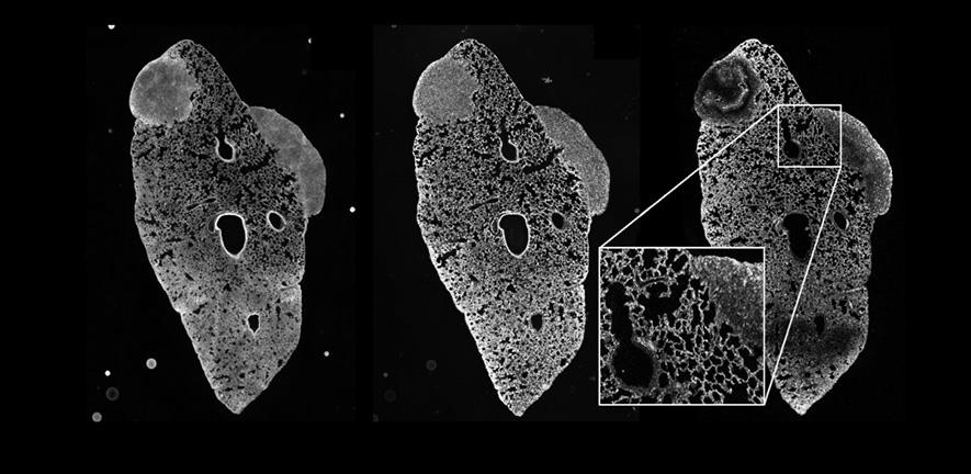
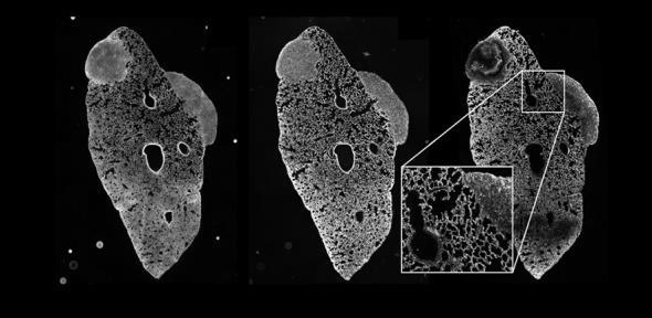
Top: Lung with two metastatic lesions derived from a mouse primary triple-negative breast tumour. The figure shows how the registration of the different imaging modalities to a cellular level allow to segment individual cells and identify tumour cell populations, differentiate hypoxic areas, increased fibrosis, infiltration of immune cells and blood and lymphatic vessels by staining with a panel of 35 cell markers at the same time. Bottom: Sample in 3D depicts a tumour grown in the mammary gland of a mouse showing the power of the SPACE pipeline to produce and visualise large volumes (typically ~100,000 individual images registered and stitched and up to 500TB 500 GB of data). The orange fluorescence beads are clearly visible in the medium outside the biological tissue and prove to be crucial for all stages of multimodal registration.
Adapted from a press release by Cancer Research UK
“The University of Cambridge is a public collegiate research university in Cambridge, England. Founded in 1209, the University of Cambridge is the third-oldest university in continuous operation.”
Please visit the firm link to site


